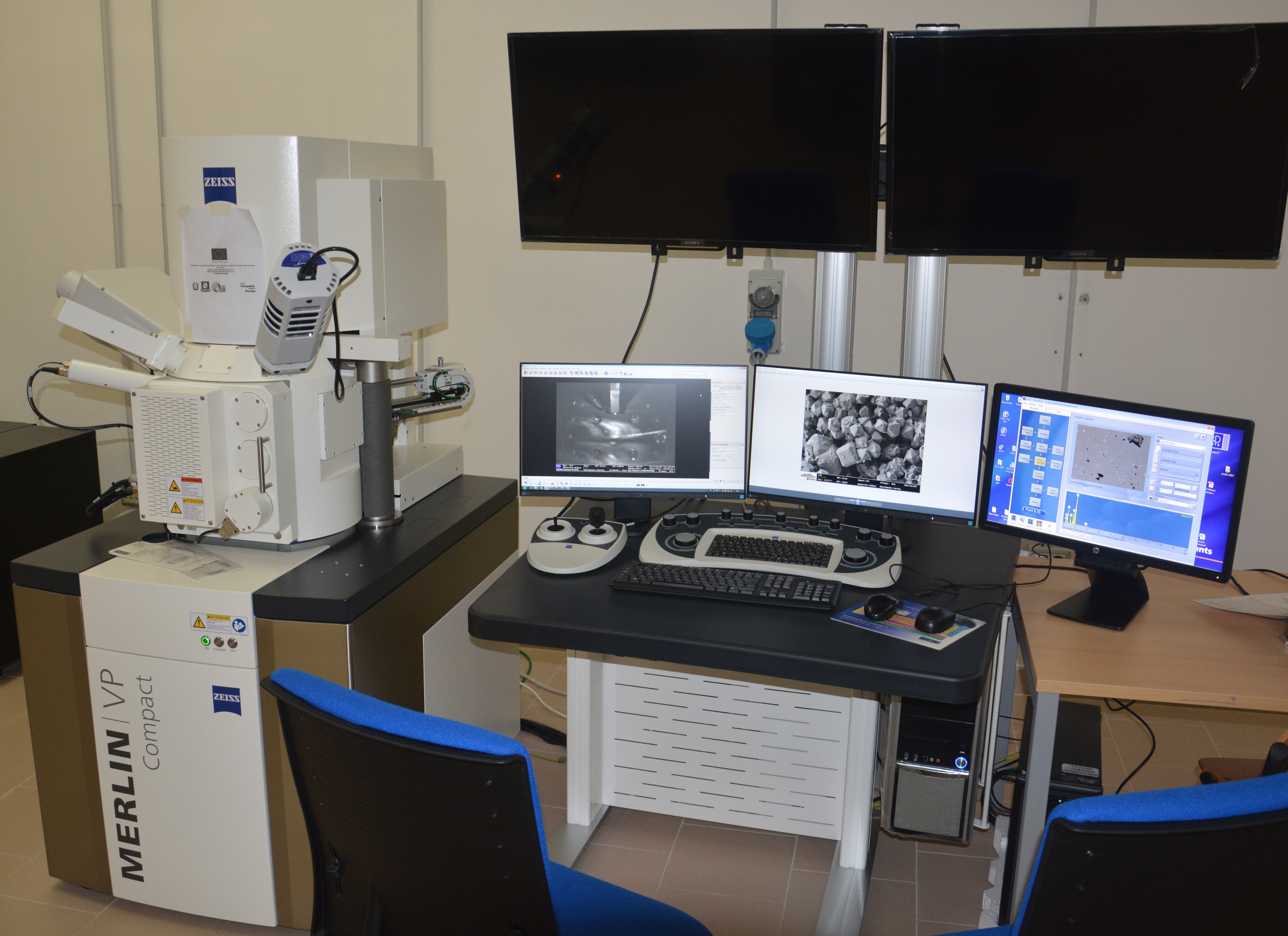
LABORATORIES AND FACILITIES

Training Courses on Safety for Equivalent Workers

Training Courses on Safety for Equivalent Workers
Safety Training for all students of the Department of Earth, Environmental, and Resource Sciences to undertake laboratory activities, geological field campaigns, educational exercises, research, and internships.
(Note: For the activation of internal internships, please pay attention to the additional provisions available at the following link.)

All university students (undergraduates, doctoral candidates, specialists, interns, scholarship holders, research fellows, and equivalent individuals) who attend educational, research, or service laboratories and are exposed to risks identified in the risk assessment document are considered equivalent to workers. As such, they are subject to preventive and protective measures to safeguard their health and safety.

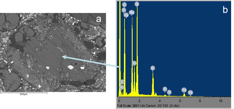
It is clarified that laboratories are considered as locations or environments where educational, research, or service activities are carried out, involving the use of machinery, equipment, plants, prototypes, or other technical tools, as well as chemical, physical, or biological agents. Additionally, locations or environments outside the constructed areas of the premises—such as archaeological, geological, or marine field campaigns—are also regarded as laboratories.

Before commencing activities involving exposure to risks, every university student (so-called "equivalent worker") is required to:

  1. Undergo a health surveillance examination.
  2. Complete the online course "Basic Training on Workplace Health and Safety" (4 hours).
  3. Attend a specific risk training course.

The Health Surveillance Examination will be requested by the tutor/professor/supervisor overseeing the laboratory activity and at the time of assigning the experimental thesis work. Notifications regarding the health surveillance examination schedule are published on the Course of Study's dedicated webpage.

The online course "Basic Training on Workplace Health and Safety" requires a 4-hour commitment (as specified in letter a) of paragraph 1 of Article 37 of Legislative Decree No. 81/08 and the State-Regions Agreement of 21/12/2011). A participation certificate is issued upon passing the final verification test.

The course is available at the following link on the Federica.eu platform:
https://www.federica.eu/partners/formazione-unina/

Access requires the use of active UNINA credentials (name.surname@studenti.unina.it).

To access the course, it is necessary to enter the access code of your department, which can be found by clicking here: https://www.unina.it/documents/11958/21142433/FORM_elenco.codici.accesso.pdf

User support and guidelines for proper course participation can be found at the following link:
https://www.unina.it/documents/11958/21142433/FORM_indicazioni.corsi.pdf

Laboratory of scanning electron microscopy (SEM)

Responsible: Prof. Paola Petrosino

Co-Responsible: Dr. Roberto De Gennaro

Tel. +39 0812538327 - This email address is being protected from spambots. You need JavaScript enabled to view it.


Laboratory of scanning electron microscopy (SEM) at DiSTAR is equipped by two scanning electron microscopes: a  SEM Jeol  JSM5310 and  FESEM ZEISS Merlin VP. Both of them allow the acquisition of high resolution magnified images of both raw inorganic and suitably prepared organic samples. Images are routinely digitally acquired.

SEM   Jeol  JSM5310, available at the lab since 1998, supplies both 3D images through a secondary electron  detector (SE ) and topographic and compositional images through a backscattered electron  detector (BSE).  Moreover, it is equipped with an EDS  X-Stream Inca Oxford detector that supplies qualitative evaluation of chemical elements starting from fluorine.  It is also possible, thanks to a well performing reference standardization, to obtain single point analyses of polished samples (resin embedded or thin sections).

Recently acquired (2017) FESEM ZEISS Merlin VP has the very large vacuum chamber Gemini 2 that hosts samples up to 15 cm diameter and its main feature is to work both in high vacuum and in variable pressure. In as much, this SEM reaches 1 million magnification. It is equipped with an "inlens" detector of both secondary and backscattered electrons, a normal secondary electron detector and a variable pressure secondary electron detector. Furthermore, it is linked to an EDS  Oxford X-Max detector and to a WDS Wave Oxford spectometer equipped with 4 analytical crystals. The combined use of EDS and WDS analytical facilities allows the quantitative evaluation of the content of chemical elements starting from boron. Dedicated softwares allow the acquisition of compositional maps for the automated evaluation of the mineralogical composition in an unknown sample.

The best advantage in using FESEM is the possibility to obtain morphoscopic and chemical data from the surface of an "as is" sample, allowing non destructive and non invasive analyses that make this apparatus the most powerful tool for SEM investigation in cultural heritage and forensic geology fields.

For SEM sample preparation, the laboratory is equipped with a gold sputter (Agar Auto Sputter Coater)  and a carbon coater (Agar Auto Carbon Coater).

The SEM lab is used for didactics, scientific research and analyses on behave of third parties. Main users are from University Federico II, other universities, CNR and other research institutions dealing with Earth Science, Physics, Chemistry, Agriculture  and Cultural Heritage. 

Our laboratory is accredited under INAIL  - Istituto Superiore di Sanità for SEM analysis of asbestos, both on total samples and on airborne fibers.

Figure 1. Field Emission Scannin Electron Microscope (SEM) Merlin VP.

Figure 2: Backscattered image of a mica crystal from a thin section of a rock (a) with the EDS spectrum (b).

Figure 3: SE image of an airborne asbesto (crocidolite) on a policarbonate filter at 2000x magnification, as the DM 6.9.1994 requires(a), and at 7500x magnification (b).

sem fig2Figura 2. Immagine in backscattering di una sezione sottile di roccia contenente un cristallo di mica (a) con in relativo spettro EDS (b).

sem fig3Figura 3. Osservazione in SE di fibre di tipo asbestiforme (crocidolite) su filtri in policarbonato a 2000x, scala dell’analisi prevista dal Decreto Ministeriale 6.9.1994 e smi (a), e a 7500x (b).

sem copertinaFigura 1. Microscopio elettronico a scansione (SEM) di tipo field emission Merlin VP.

logoUniNa   
scuola psb
universitaly

Where we are

  whatsapp

Find us

  Find us on Google Map

Newsletter subscription